
FCP portfolio case study
The management of a paediatric patient with metatarsalgia demonstrating clinical excellence by first contact podiatrist.
Case study
FCP portfolio case study
The management of a paediatric patient with metatarsalgia demonstrating clinical excellence by a first contact podiatrist
A case of Freiberg’s disease in a nine-year-old girl, early detection, and management
Previous history
A nine-year-old girl was booked into the out-of-hours first contact podiatry telephone consultation. This was done following a review with the GP who noted in the patient's record there was generalised pain in the second and third toe for several weeks, normal range of motion, no tenderness when assessed and examined, no clinical signs of infection and unable to find cause of pain and wanted a second opinion.
Following a telephone consult a face-to-face was offered the following day. Although I was not concerned about any red flags from our conversation, a physical assessment was deemed necessary.
Clinical assessment and presentation
Presentation of unresolved pain in the front of the foot and the patient had been walking on the side to avoid the painful area. The pain was unremitting, described as a dull ache and located only to the right foot with an impact on toe push off. The duration was three months with no history of trauma or injury. There was no hand pain noted, no sudden weight loss and no early morning stiffness reported.
Mum thought it was growing pains but was now concerned as it was affecting gait. The patient was in good health, systemically well and was engaging throughout the consultation. She reported no pain at night.
On assessment, there was bilateral syndactyly of the second and third toes. Examining the joints for a range of motion there was definite reduced dorsiflexion and plantarflexion of the second digit at the second metatarso-phalangeal joint (MTPJ) compared to the left foot.
Gait showed no abnormalities apart from reduced propulsion through the second metatarsal head to the right foot. Normal muscle strength and power were evident and unremarkable. There were no neurological concerns with 100% detection of 10g monofilament and normal reflexes. Pulses were palpable. There was no oedema and no erythema noted on the digits. The impact on life was that she was managing school, but PE was becoming difficult and the pain not improving.
Investigations
Based on the clinical findings I referred the patient for ultrasound and x-ray due to the reduced range of motion in the second metatarso-phalangeal joint.
Differential diagnoses and clinical reasoning
Differential diagnosis based on this presentation included fracture, Morton's neuroma, or Freiberg's. For me recognising what was a normal range of motion and what was acceptable was the most important aspect of the assessment and the type of pain that was being described. With the duration being three months, absent of trauma it was unlikely to be a fracture unless it was a stress fracture. Equally, both Morton's neuroma and Freiberg's can cause pain around this area.
The pain associated was not considered to be pins and needles nor was it a burning sensation - more like a dull ache, and no mention of a stone-like feeling on the ball of the foot nor like walking on a pebble.
Freiberg's was my initial preferred diagnosis, but I felt she may be too young. Though Love and O'Mara in 2010 reported it is found in all age groups from children to the elderly, Freiberg's most commonly affects adolescent females, with a 5:1 predominance over males. This is the only osteochondrosis that favours females more than males (Cerrato 2011). On examination, the patient demonstrated a reduced range of motion and pain around the second metatarsal head area, with no indication of injury and trauma so did have the feature of Freiberg's.
In referring for an X-ray this would rule out any fracture or any bony abnormality. If suspecting early Freiberg's, then an X-ray can often miss the features and the patient is often then misdiagnosed or discharged. Ultrasound can be much more effective at picking up earlier, subtle changes.
An ultrasound scan of the area was undertaken and an X-ray. The patient was fitted with an off-loading device and appropriate follow up arranged with a safety net in case of any issues.
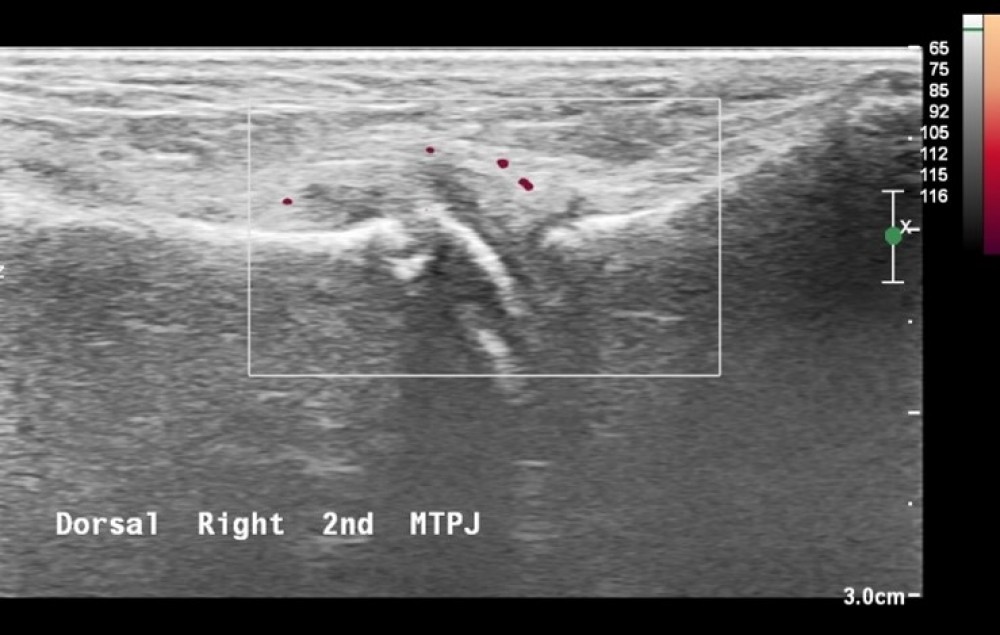
Image: Ultrasound of right forefoot showing increased vascularity to the dorsal second MTPJ, joint effusion and distension.
The ultrasound scan of the patient's foot shows joint effusion around the second MTPJ and a distended joint capsule. The report suggests some periosteal surface irregularity on the neck of the metatarsal – which could be deemed as age-related ossification, however, is relevant to the other pathology and the clinical symptoms.
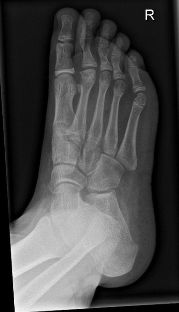
Image: X-ray shows Freiberg's disease with flattening of the second metatarsal head
Early detection
Early detection and treatment of this pathology are needed. This allows for the appropriate conservative treatment to be delivered to prevent needing surgery, reduce pain levels for the patient, and improve outcomes.
Presenting symptoms can be minimal with problems developing later in life if not managed appropriately. It can vary in severity and may develop over time. Complex cases can cause the bone to breakdown, causing early arthritis in the joint and leading to long-term problems, such as pain, unable to wear certain footwear, and participate in activities.
What is essential is early detection of this condition to allow for appropriate treatment options to prevent long-term effects on the foot.
Reference
Cerrato, R.A. (2011). Freiberg’s Disease. Foot and Ankle Clinics, 16(4), pp.647–658. doi:10.1016/j.fcl.2011.08.008.
Love, J.N. and O’Mara, S. (2010). Freiberg’s Disease in the Emergency Department. The Journal of Emergency Medicine, 38(4), pp.e23–e25. doi:10.1016/j.jemermed.2007.10.028.